22.09.2020
Mole under the toenail. Treatment of subungual melanoma at the initial stage. Possible complications and consequences
Cancer is one of the greatest problems of humanity, which is becoming more and more urgent every year. A tumor can appear anywhere in the human body and, during its development, spread to large areas by metastasis. Skin cancer (and it also has several types) is one of the most common pathologies in oncological practice. But the primacy in mortality in the first year of the disease is still held by one of the types of skin cancer - melanoma. True, melanoma is often understood as a tumor of melanocytes on the surface of open skin areas, and not everyone suspects that such a neoplasm is possible in the nail area. Dark spots under the nail are often attributed to an injury with hemorrhage (bruise, hematoma), but in fact it may be a malignant formation - subungual melanoma.
, , ,
ICD-10 code
C43.3 Malignant melanoma of other and unspecified parts of face
Epidemiology
Since cell degeneration occurs when cells are damaged, the risk of developing nail melanoma is relatively low. Still, the cells of the nail bed are protected from damage by the dense nail plate. According to statistics, only 0.7-4% of tumor processes on the skin are diagnosed in the nail area.
At the same time, the risk of developing subungual melanoma on the hands or feet is approximately the same, which cannot be said about different fingers of the extremities. The big toe is most susceptible to injury (especially on the feet), so melanoma of this toe is most common. By the way, in 4 out of 10 cases of nail melanoma, patients point to its injury in the recent past.
Most often, the disease affects adults. After 55-60 years, this figure is maximum. Subungual melanoma is unlikely in children. Usually, a dark spot in the area of \u200b\u200bthe child's nail turns out to be a nevus, causing the appearance of a characteristic strip (melanonychia) on its surface.
People with dark skin color (African Americans, Indians, Hispanics, Asians) are most susceptible to the development of subungual melanoma.
In representatives of the dark-skinned race, the disease develops mainly against the background of melanonychia (deposition of melanin in the nail plate). The dark-skinned inhabitants of the planet have a tendency to form dark spots on the nail bed and in the nail plate, but often pathology is not considered as an independent disease, considering it a symptom of other diseases, including subungual melanoma.
, , , , , , , , ,
Causes of nail melanoma
We have identified several factors that can lead to the degeneration of cells in a certain area of \u200b\u200bthe skin: trauma, UV radiation, pigmented nevi, hereditary predisposition. Now we will try to consider the causes of melanoma under the nail more extensively.
To begin with, speaking of nevi, we mostly suspect moles or birthmarks. In fact, hemangiomas (not a malignant vascular tumor, which is usually detected immediately after the birth of a child), papillomas (a benign tumor, which is believed to be caused by a papillomavirus), and warts (viral neoplasms on the body) also have similar properties. Despite the fact that all such growths are benign, their damage is likely to lead to a change in the properties of cells and malignancy of the process.
In principle, the cells of any growth on the skin of a finger, which has existed for many years, without bringing the owner much anxiety, in case of injury, take the brunt of the blow, and therefore are damaged more than others. At the same time, the risk of developing malignant processes in this case is higher, and it does not matter where this growth was formed: on the open skin or under the nail plate.
After the age of 40, some people develop separate shapeless dark spots on the skin that resemble moles. At the age of over 50, such marks appear already in many and not one by one. This pathology is called seborrheic keratosis (senile keratoma), and it is caused by a violation of the production of keratinocytes in the basal layer of the skin. The neoplasm itself is benign. But over time, it begins to rise more above the skin and becomes more prone to injury. If such a stain appears on the toes, it can be squeezed or rubbed with shoes, suffer during impacts, etc., which can lead to cell degeneration. And there are quite a few such cases.
Risk factors
Risk factors for the development of subungual melanoma are any previously diagnosed types of skin and connective tissue cancer, as well as a hereditary predisposition to cancer. Even if the cancer is localized inside the body, it can metastasize to the area of \u200b\u200bthe nail fold, base of the nail, etc.
When we talked about dark-skinned people, we noted that a predisposition to subungual melanoma in them is associated with more frequent cases of melanichia. The incidence of this disease in Caucasians is less than 1%. But this does not exclude the appearance of a subungual type of melanoma in people with fair skin. Regardless of the localization of the lesion focus, melanoma is more susceptible to people with fair skin (usually they have light or red hair and blue eyes), the presence of a large number of moles, with freckles on the face.
It is clear that the risk of malignant cell degeneration is higher in people who like to take sun baths, especially during the hours of increased sun activity, sunbathe in solariums, and work in the open air. As for solar radiation, often skin burns received in tender childhood become an oncological problem in an adult, sometimes after several decades.
Fingers are areas of the limbs that are injured very often. But if even in domestic conditions injuries to fingers and nails are not a rare occurrence, then what can we say about production conditions, where the bulk of the work is done by hand, or playing sports with a high risk of toe injuries (for example, football) and the development of big toe melanoma , because it is this finger that suffers most often and more than others.
It is clear that without provoking factors, no growth on the fingers or under the nail plate will turn into a malignant tumor. But how to avoid these provoking factors, if our living conditions, nutrition, work already pose a risk of injury, poisoning of the body with carcinogenic substances, exposure to sunlight UV light. It turns out that the risk of developing subungual melanoma is quite high for each of us living in modern conditions, working in production and forgetting the taste of natural products. The villagers benefit from this.
Pathogenesis
Symptoms of nail melanoma
Subungual melanoma is a disease similar in symptoms to some other diseases: nail fungus, hematoma after injury, subungual nevus, melanonychia, wart under the nail, paronychia or panaritium (inflammation or pus formation in the area of \u200b\u200bthe nail fold and base of the nail). This is what makes the diagnosis of pathology difficult.
And yet, what should alert a person? What signs can indicate that the appearance of a dark spot and inflammation in the nail area is not a simple consequence of an injury, but the beginning of a malignant process? Consider the symptoms characteristic of the onset of the disease:
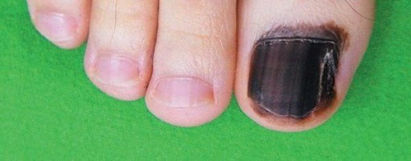
- The first sign of a possible pathology is a change in the color of the nail and the tissues under it or around the nail plate. It does not have to be the entire nail, most often the discoloration is observed in some part of the nail, for example, at its base. In this case, fabrics can be painted in burgundy, rich, red, brown, violet-black and even blue.
It is clear that darkening of the tissues in the nail area is possible due to severe trauma with hemorrhages (bruising). Trauma symptoms usually resolve within 10-12 days. If this does not happen, you should consult a doctor and diagnose the site of damage.
Naturally, if the dark spot was not formed due to injury, it must be examined.
- If we are talking about non-pigmented melanoma, then darkening of the tissues may not be observed. And there is no pain in the early stages of the disease. For this reason, the non-pigmented form of the disease is usually diagnosed with a delay, when its treatment is already very difficult and rarely brings good results.
But both pigmented and non-pigmented melanoma grows little by little and a longitudinal stripe appears on the nail plate above it. Most often, such a strip, which has a darker color compared to other tissues, is located strictly in the middle of the nail, but it happens that it is shifted to the right or left from the center of the nail plate. An identical situation is observed with melanonychia.
Over time, the stripe becomes darker and wider. In this it differs from the strip on the nail plate caused by hemorrhage due to microtrauma or taking certain medications, which does not change in size over time and shifts as the nail grows. In the dark-skinned race, the appearance of a dark stripe is a certain feature, and does not always indicate melanoma.
The melanoma strip expands until it covers the entire surface of the nail. Sometimes the process spreads to the lateral (lateral) nail folds, which also change the skin color to a darker one.
- At first, the tumor is not palpable and it can only be suspected by a change in the color of the nail tissue, but as it grows, it thickens and begins to press on the nail plate, causing its destruction. The nail exfoliates, becomes brittle, cracks appear on it. The adhesion to the nail bed decreases. All of these symptoms are very similar to nail fungus.
As the disease progresses, blood and pus begin to ooze from under the nail bed. The tissues around the nail become inflamed, and abscesses form between the nail plate and lateral nail ridges. From the outside, it looks like a common inflammation of the periungual fold (paronychia). With the appearance of suppuration in this area, felon can be suspected. But in fact, everything can turn out to be much more dangerous, because the appearance of pus from under the nail and in the area of \u200b\u200badjacent tissues is one of the symptoms of subungual melanoma.
Further, ulcers form at the site of the abscesses, which gradually become larger in size. Attempts to treat the disease with anti-inflammatory and antibacterial ointments do not work, because we are not talking about an infectious process. The sores may fester or ooze, and they are quite painful, but will not heal, no matter what measures are taken.
If at first the melanoma resembles a small tubercle, then over time it changes its shape to mushroom with a fleshy "cap" and a thinner leg. This is a characteristic sign of melanoma, although again there is a similarity with papilloma.
It must be understood that the malignant process tends to spread not only over the surface of the skin, but also inside the tissues. If at the first stage there is practically no pain when pressing on the nail, then with the spread of the tumor process to other areas and deep into the bone, the pressure on the finger will be accompanied by severe pain. When pus appears under the nail, the pain can be constant, pulsating.
It is clear that inflammation with the formation of pus and dystrophic processes in the nail disrupt the nutrition of the nail plate, as a result of which it is separated from the nail bed, on which the malignant process is actively progressing. But now he is no longer hidden from view, and there is no doubt about his character. True, treatment at this stage of the disease has not so encouraging results.
Stages
Melanoma symptoms may vary somewhat depending on the type of pathology and its stage. There is no strict classification by type regarding nail melanoma, but they are distinguished:
- melanoma, which develops in the area of \u200b\u200bthe matrix (base) of the nail, then darkening of the nail is immediately observed in the area of \u200b\u200bthe lunula,
- melanoma that originates under the nail plate (in this case, a dark spot can appear anywhere on the nail and stretch into a colored strip as the nail grows),
- melanoma of the skin near the nail plate (the spot appears on the periphery of the nail, but gradually spreads further).
It should be understood that all of these features apply to pigmentary melanoma. With a pigmented form, external manifestations are not observed until brittle nails, pus and ulcers appear. Also, over time, you can feel the seal under the nail.
As for the progression of the disease, the initial stage of subungual melanoma is more reminiscent of a subungual hematoma with the appearance of a dark spot on the nail, which gradually lengthens and grows with the nail. There are usually no other signs.
In the second stage of the disease, the nail is destroyed, and purulent inflammation appears. By the end of the second stage, multiple ulcers are observed under the nail plate and next to it, from which the ichor oozes. Further delamination of the nail occurs.
It is known about the third and fourth stages of subungual melanoma that during this period the process of metastasis occurs. First, there is inflammation of the regional lymph nodes and their compaction due to the proliferation of malignant cells and individual foci of the tumor process in nearby tissues, subsequently, distant metastases appear that affect the internal organs of a person.
Depending on how the malignant cells spread: with the flow of lymph or through the blood (lymphogenous and hematogenous pathways of metastasis), the disease will proceed slowly (in the first case) or aggressively, in a short time affecting large areas of the body (when spreading with the blood flow through the blood vessels).
, , ,
Complications and consequences
A cancerous tumor is a terrible neoplasm, regardless of where it is found. After all, cancer cells not only multiply excessively, forming seals that compress nearby organs and disrupt their functionality. They also poison the body and destroy healthy cells. Through their fault, various failures occur in the body, and when it comes to vital organs, the patient's death occurs.
Usually death is associated with a large tumor size or metastases in the heart, kidneys, lungs. As long as the tumor is small and has not metastasized, it can be safely removed, but again it all depends on the extent of the process. Sometimes surgeons are limited to only removing the nail or distal phalanx of the finger, in other cases it is necessary to remove the entire finger. If the process of metastasis has not been started, one can hope for a favorable outcome of treatment, otherwise malignant foci may subsequently appear in different parts of the body.
In the case of the spread of malignant cells through the blood, the disease develops very quickly, and by the time it is diagnosed it can go into the third or fourth stage. First, a person suspects a nail injury with the formation of a hematoma, then begins to treat the destruction of the nail and abscesses, considering them the intrigues of fungi and bacteria, and when the treatment does not work, he comes to the doctor, who diagnoses the advanced stage of the disease, although it was possible only a couple of months after the appearance first symptoms.
The situation is not the best with non-pigmented subungual melanoma. At first, she does not show herself in any way, then such a light wart appears, lifting the nail. Over time, the wart takes on a specific mushroom shape, and it would be time to think about what it could be. But until it hurts, few people begin to worry about her appearance. When pains appear, it turns out that the process has gone deep inside and affects the bones.
Diagnosis of nail melanoma
If you carefully study the symptoms of subungual melanoma, you will notice that there are actually no specific symptoms by which the disease could be diagnosed with great accuracy. The neoplasm at different stages of development will resemble the clinical picture of many other diseases, which significantly complicates the diagnosis. And often it simply delays the moment of going to the doctor and becomes the reason for delayed treatment without any guarantees.
By the appearance of pigmented melanoma, the doctor can only suspect oncology. To confirm the diagnosis, you will also need to prescribe a clinical blood test and a blood test for tumor markers. But again, a positive result indicates only the presence of a malignant process in the body and does not provide information about its localization. Perhaps the patient has a common hematoma on the nail, which will be removed by mistake, but this will not relieve him of a tumor elsewhere, which no one even suspected.
It is possible to confirm that a dark spot on the nail is melanoma using instrumental diagnostics. Of course, dermatoscopy comes to the fore, i.e. examination of the spot under the nail using a special device - a dermatoscope. This modified microscope allows you to see through even the stratum corneum of the nail plate in order to detect malignant cells under it or to exclude the diagnosis of cancer.
Digital dermatoscopy allows you to get an enlarged image of damaged tissues on a computer monitor and make a high-quality image for further study and consultation.
It is clear that at an early stage, when there are still few malignant cells, dermatoscopy may not reveal them, especially if the study is carried out by a doctor with insufficient experience. The only way to 100% confirm or deny the diagnosis of subungual melanoma is a biopsy, during which a tissue sample is taken from under the nail to a depth of 3 mm.
To get an informative sample in the area of \u200b\u200ba dark spot at the base of the nail, you need to puncture or remove the nail plate. But it is best to remove not only the nail, but a suspicious neoplasm with adjacent healthy skin areas. The fact is that a biopsy, i.e. violation of tumor tissues can provoke even greater cell proliferation and tumor growth, which, moreover, can give metastases in a short time. In order not to risk the biopsy, it is necessary to take a biopsy from a remote area of \u200b\u200bthe nail, which includes the nail plate, muscle tissue under it, subcutaneous fat, and sometimes the entire distal phalanx of the finger, if the process was widespread.
There is no need to worry about the removed nail and the wound in the nail bed. The wound will heal, perhaps even a new nail will soon grow back. But it is better to remove the hematoma than to ignore the cancer or disturb it and cause metastasis. At an early stage of the disease, the probability of a complete cure is quite high.
If a biopsy confirms the diagnosis, it is important to understand at what stage the disease was detected and whether the tumor has metastasized. To detect metastases, they again resort to instrumental diagnostic methods: ultrasound examination, radiography, computed and magnetic resonance imaging. If there is an increase in regional lymph nodes, a puncture biopsy is required to determine if the change in the size of the lymphatic vessels is associated with the proliferation of malignant cells inside them.
,,, [due to the similarity of external signs and the nature of the lesion. A subungual wart may raise suspicion of a non-pigmented melanoma, and a subungual hematoma may raise a suspicion of pigmented melanoma.
In order to differentiate diseases and exclude an incorrect diagnosis, it is very important, according to the patient, to study the behavior of a spot or tubercle in the nail area. With melanoma, they will certainly increase in size. Pay attention to the nature of the pain. The appearance of a dark spot that is not associated with trauma and does not cause pain when pressed is most likely to indicate its malignant nature.
Treatment of nail melanoma
As we have already said, cancer cells are highly viable, so chemotherapy or radiation without removing the tumor can hardly guarantee the complete destruction of the malignant particles. In addition, in order to accurately diagnose cancer based on the results of a biopsy, it is still more expedient and safer to remove the neoplasm first. It turns out that the surgical removal of subungual melanoma is the main method of its treatment.
If the tumor is relatively small and has not gone deep into the roots, the phalanx is sanitized with the removal of the nail plate and soft tissues under it to the depth of the tumor penetration plus some part of healthy tissue is captured. If the process has spread to the periungual ridges, even part of the finger bundles is removed, but the phalanx is preserved.
In the case of deep penetration of the tumor and metastasis into the bone tissue, sanitation, as a method of surgical treatment, no longer makes sense and it is necessary to resort to amputation of the distal phalanx (less often of the finger). Thus, the hematogenous spread of cancer cells can be prevented.
If enlarged lymph nodes are found after a biopsy and confirmation of the presence of cancer cells in them, lymphadenectomy is prescribed, i.e. excision of the lymph node affected by oncology. All this is done in order to block the lymphogenous pathway for the spread of cancer.
Other treatments for nail melanoma are prescribed only after the tumor has been removed. We are talking about chemotherapy (drug treatment with potent agents), radiation treatment, which is a dosed irradiation of a certain part of the body in the area of \u200b\u200bthe finger (if the tumor has metastasized, other parts of the body are also irradiated), taking immunomodulatory vaccines and serums containing antibodies. Immunotherapy is effective for melanoma.
With regard to chemotherapy, treatment can be carried out with two types of drugs - cytostatics and cytotoxic drugs. The former inhibit the reproduction of cells and lead to their decay (death) due to the inability to divide, the latter lead to the intoxication of cancer cells, as a result of which they die.
After removal of the tumor without metastases, local treatment with chemotherapy can be carried out (application of ointments, washing the wound with solutions, injecting drugs directly into the tissues of the affected finger). If metastases do not extend beyond the finger, regional chemotherapy is performed, i.e. drugs are injected directly into the diseased organ. When metastases spread beyond the finger and spread to internal organs, systemic chemotherapy is required (drugs are administered intravenously or orally).
Usually, even such a complex and difficult to tolerate treatment gives good results only in the early stages of the development of the disease. Further, it only slightly prolongs the life of patients and requires additional administration of anesthetic drugs, since the spread of the malignant process inside is always accompanied by severe pain.
Prevention
Oncologists believe that you need to pay close attention to any changes in the nail after an injury. Such changes should be considered malignant until proven otherwise. It may seem strange to some, but such an attitude to the problem is justified. Better to be safe than to die because of a running bruise, which at some point turned into a cancerous tumor.
When talking about the treatment of cancer, it rarely comes to long-term forecasts, because it is not always possible to get rid of cancer cells completely, besides, the disease greatly depletes the body and makes it more susceptible to other diseases, for example, diseases of an infectious nature, against which the forces no longer remains. Not only the disease weakens the body, but also chemotherapy or radiation therapy. Sometimes the tumor forms again, but in a different place.
Usually, when making any predictions, they talk about a five-year survival threshold. It is believed that if the patient does not die during this time, i.e. the disease has not returned, there are chances for a full recovery.
So at the initial stage of subungual melanoma, the five-year survival rate is 75-88%, which is considered a high indicator. The forecast for the second stage is already much worse - about 60-70%. At the third stage, when metastases spread only to the lymph nodes, we can speak only about 40%, and then there is a great risk of relapse 5 years after the operation with a sluggish course of the process. When internal organs are damaged (stage 4), up to 85% of patients die within five years, and only a few cross this threshold.
It turns out that the sooner a patient seeks help, the more chances he has for a full recovery with the appointment of adequate therapy. This suggests that you need to pay attention to any changes in the color or shape of the nail, the appearance of new nevi and seals on the fingers, the formation of longitudinal lines on the nail and their behavior. If there is a change in the color of the subungual nevus, an expansion of the strip on the nail and a thickening of the nail plate, it is imperative to consult a doctor.
The only thing that remains for us is to carefully monitor our health, be as careful and careful as possible, regularly examine our body for the presence of incomprehensible growths, bumps, age spots, pay attention to changes in the color and shape of moles, the appearance of dark blotches on them, ulcers, cracks, changes in the shape and thickness of the nail. Only in this case there is a great chance to avoid such a deadly disease as subungual melanoma. Do not think that if the pathology is rare, then it will not affect us. This attitude to the problem is fraught with great disappointment.
It is important to know!
Today, various neoplasms that affect the skin are increasingly common. Moreover, about 4-10% of them are malignant tumors. People of different genders are affected with the same frequency. In most cases, the tumor does not form spontaneously.
This is the name of the malignant degeneration of pigment cells - melanocytes. It is known that the disease can affect the skin. However, there are cases when abnormal cells can form under the nail.
The disease is quite rare. It occurs somewhat more often in men than in women. The growth of such a tumor occurs much faster than other malignant neoplasms, which explains the high mortality rate from this disease. Melanoma of the nail has several varieties:
- A tumor that grows out of the nail matrix.
- Melanoma that forms under the nail plate.
- A tumor that grows on the skin next to the nail plate.
Melanoma causes
Melanoma of the skin in open areas, as a rule, develops at the site of a mole or nevus under the influence of ultraviolet radiation. Experts noted several factors that could affect the transformation of cells into malignant ones:
- Nail trauma, even long-standing. It can be bruised, burned, squeezed, scrapped.
- Genetic predisposition to pathology.
- Frequent exposure to sunlight or UV radiation in a solarium.
- Elderly age.
- The presence of bad habits and weak immunity.
More susceptible to the development of melanoma under the nail are fair-skinned, red-haired people and people with a large number of birthmarks and age spots, moles and freckles.
The tumor can be localized under the nail plate, affect the nail fold, or form on the skin next to the nail. Outwardly, the disease looks like a dark spot on the skin with uneven edges and irregular shape.
It is often confused with panaritium or hematoma. The color of the formation varies from light brown to dark brown.
You can also observe shades of burgundy and purple.
Age, gender, or skin color doesn't matter for melanoma. This disease can affect anyone. That is why it is so important to know the reasons that contribute to the development of melanoma. Experts do not yet know the exact reasons provoking the development of the disease. The factors that increase the chances of getting sick are:

The reliable cause of the development of melanoma has not been established. But doctors point to the existence of risk groups, among which skin cancer is more common. This category includes people with the following characteristics:
Absolutely everyone can get nail oncology, regardless of age, skin color, race and other characteristics.
Etiology and types
Melanoma is an atypical degeneration of melanocytes or pigment cells. The disease affects the human dermis, but, in rare cases, abnormal cells can appear under the nails.
The disease is considered rare; men suffer from it more often than women. Subungual melanoma is characterized by rapid growth and high mortality.

Melanoma of the nail is divided into several types and categories.
Types of disease:
- formation that affects the nail matrix;
- a tumor localized under the nail plate;
- melanoma on the skin, next to the nail plate.
All of these species pose a danger to human life. In terms of the number of deaths, the disease is the leader among skin cancers.
Melanoma can develop from the nail matrix or originate on the skin near the nail plate. There is also acral nail melanoma or melanonychia. It appears on his bed and is a dark line located longitudinally. It is often mistaken for an ordinary bruise and is not sought for help until it is too late. Symptoms to watch out for:
 Melanoma can be located under the nail or near its horny part.
 - the nail plate quickly changes color;
- the nail is undergoing changes;
- when pressing on the plate, pain appears;
- a crack appears along the nail.
Depending on the localization of the malignant process, the following cancers of the nails are distinguished:
- Melanoma of the nail plate.
- In fact, subungual melanoma.
- Melonoma, the development of which occurs from adjacent tissues.
Possible stages
As mentioned above, there are 4 stages of nail melanoma. Let us consider in more detail the signs and characteristics of each phase of the disease.
Melanoma under the nail develops gradually. At first, this disease may be completely invisible or "disguised" as another problem. Often, patients go to the doctor when it is too late - at the third or fourth stage of the disease. There are four stages of cancer development.
The presence of moles and age spots requires a person to regularly monitor their condition. If abnormal changes in the structure of the nevus are found, it is necessary to immediately consult an oncologist.
In oncological practice, two main methods of therapy are used to treat melanoma in the early stages:
- A surgical procedure that removes all cancerous tissue.
- A complex technique, including surgery and radiation therapy. The combination of surgical excision of the tumor and radiological technique gives the most favorable results and the chances of cancer survival... Radiation exposure through the use of highly active X-ray radiation allows you to destroy possible cancer cells in the lymph nodes.
Radiological treatment is used in the form of remote irradiation of the tumor before surgery. This is necessary to stabilize the malignant process and prevent recurrence of the disease.
Patients with stage 1 oncology have a positive prognosis of five-year survival - more than 80%. Stage 2 cancer reduces survival by up to 55%. What do these indicators mean?
For example, a survival rate of 80% means that after treatment, 80% of patients survived 5 years or more without relapses and complications. The third stage of nail melanoma with metastases to the lymph nodes reduces the prognosis to 30-40%. At stage 4, the prognosis is the least favorable - only 15%.
Modern medicine can successfully fight melanoma in the early stages of the development of the disease. Therefore, pay close attention to your body and regularly examine your body.
If you notice pigmentary defects on it that change size or shape, then immediately contact a dermatologist or oncologist.
Signs of nail melanoma
Symptoms of subungual melanoma change with the course and progression of the disease. Therefore, it is important to notice its first signs in time. Namely:
- The appearance of a small pigment spot that has formed under the nail plate. The spot can be in the form of a small longitudinal strip on the nail bed. Sometimes the appearance of subungual melanoma is preceded by a small finger injury, due to which the patient did not consult a doctor in a timely manner.
- An increase in the stain under the nail (usually within a few weeks or months). It becomes wider, especially in the cuticle area, changes color to dark or light brown.
- The spread of education on the nail roller.
- The development of nodules and the appearance of bleeding ulcers, leading to thinning, cracking and deformation of the nail plate (nail dystrophy).
As the malignancy develops, the entire nail is affected.
The presented type of melanoma accounts for 10% of the total number of tumors. Neoplasms occur with the same frequency in people of any race (you can see how it looks on the skin in the photo). Moles do not precede the development of the lesion.
If melanoma is noted on the nail bed, its sign is the presence of a brown longitudinal line. The tumor cannot be felt by touch until it enters the stage of vertical growth.
The nail plate begins to rise above the finger, more of the symptoms should be noted:
- persistent paronychia;
- painful sensations;
- dystrophy of the nail plate;
- increased pigmentation;
- longitudinal splitting of the nail.
Symptoms of nail cancer
Some people are worried about whether an ordinary mole can become a tumor. In some cases, this is the case. Often a person notices that a mole has appeared on the skin near the nail. It could be melanoma.

Cancer of the nail has striking characteristic features. The initial stages of the disease often occur without pronounced clinical symptoms, which complicates early diagnosis and timely treatment.
If you notice a characteristic darkening of the nail plate or the appearance of a dark area near it, rush to the doctor: this change may indicate that a cancerous tumor of the nail is developing in the body.
The common symptoms of nail cancer are as follows:
- The appearance of a characteristic dark or black spot on the nail. It grows very quickly and sometimes splits the nail exactly in half.
- Darkening of the nail bed (this is the first and earliest sign of malignant nail cancer).
- Cancer covering the entire nail plate (this rarely happens at stage 1, it usually happens in a few months).
- Be careful: sometimes formations appear on the nail plate, not only black, but also blue, brown and even purple.
- If you damage the nail plate, then bleeding is possible.
- The nail plate is eroded and destroyed due to ulcers.
- Purulent contents are released from under the nail plate.
Melanoma in the foot can spread to the sole. This process leads to severe walking difficulties. Sometimes a person cannot stand on the affected leg at all.
The disease is quite rare. Slightly more common in men than women
Characteristics of this melanoma
The early stages of this disease are quite insidious. This is due to the fact that the doctor may confuse them with hematoma or subungual panaritium.
The further course of the disease causes the appearance of a mushroom formation on the nail plate. Finally, the nail is completely affected and flakes off.
Such phenomena indicate a neglected disease. Treatment of advanced forms of this disease is very difficult.
The rate of development of nail plate melanoma can vary significantly. With the slow progression of cancer, metastases appear at the very late stages.
There is also a fulminant form of melanoma, when metastases with blood flow extremely quickly spread to distant organs. Usually, palliative treatment is prescribed for such patients.
The following symptoms indicate tumor metastasis:
- the appearance of a tangible seal under the nail plate;
- the presence of a chronic cough;
- change in skin tone (it becomes dull, ashy);
- the patient's body temperature rises;
- the nail is almost completely destroyed and bleeds;
- seizures develop;
- lymph nodes increase and thicken;
- weight loss (sometimes even to cachexia).
What is melanoma and how to detect it yourself (video)
How the disease progresses
Subungual melanoma has its own specific features by which it can be distinguished from other pathologies. As the disease progresses, symptoms begin to change, and a minor defect becomes a visible neoplasm.
- a small pigment spot or strip under the nail plate;
- the defect does not go away, unlike a hematoma after two weeks;
- the age spot increases in size;
- a color change occurs up to black or dark brown;
- the edges of the defect begin to bend;
- education moves to the nail roller;
- cracks, bleeding wounds, nodules appear on the roller;
- the nail plate is deformed.
Important! Subungual melanoma does not always change color. The disease can develop for a long time, without any symptoms. There are cases when nail melanoma spread to the sole.
In the initial stages, this pathology is very insidious. This is due to the fact that the disease can be confused with the usual hematoma or nail felon.
The tumor growth rate changes frequently. With slow development, metastasis occurs at the last stage of the pathology, and with rapid progression, metastases are spread by blood to distant organs.
The process of metastasis can be determined by the following signs:
- a noticeable seal under the nail;
- chronic cough;
- skin color becomes gray, dull;
- high body temperature;
- the nail collapses and begins to bleed;
- convulsions appear;
- lymph nodes increase;
- the patient's weight is sharply reduced.
Most often, this disease is found in the area of \u200b\u200bthe big toe or toes - it is usually deployed not on the nail, but on the skin under or next to it. Gradually, the disease develops, spreading throughout the nail plate. You need to sound the alarm in the following cases:
- a dark spot grows on the nail plate (it darkens completely in a month or two);
- painful sensations appear when pressed;
- hematoma under the nail does not go away within 14 days;
- bleeding in the nail area, the appearance of pus;
- color: red, purple, brown and up to purple and black;
- sores under the nails.
The primary manifestations of subungual melanoma are virtually absent, and only during the development of a cancerous lesion is the external manifestations of the tumor detected in the form of:
- The presence of a small brown spot that is located at the base of the nail bed. In some cases, the nail lesion may develop as a longitudinal strip on the nail. From time to time, before the origin of external manifestations, patients note a finger injury, for which timely medical care did not appear.
- In the later stages of the disease, the age spots increase in size and the color becomes much more intense.
- During growth, a malignant neoplasm spreads to the entire nail plate and adjacent soft tissues.
- The terminal stages of the disease are accompanied by the formation of a nodular lesion of the nail tissue, followed by ulceration and the origin of spontaneous bleeding.
Considering the rate of development and growth of this malignant skin lesion, early detection of the tumor is very important for successful treatment. Accurate diagnosis is difficult because melanoma is easily confused with a common nail injury.
At the initial stage, this malignant formation is often mistaken for a hematoma after injury, paronychia (suppuration) or panaritium (inflammation of the periungual ridge).
Therefore, it is necessary to know the main symptoms of this dangerous disease:
Most often, melanoma is found on the large nails of the hands or feet. A swelling on the toenail is more dangerous. The neoplasm does not develop on the nail plate, but under it or next to it in the skin.
While developing, it spreads to the entire nail plate. The main symptoms of melanoma are as follows:
- a dark spot appears on the nail, gradually increasing in size;
- the appearance of the spot may be preceded by trauma (if there is a hematoma under the nail, then it will pass within 2 weeks, if it does not go away, then you need to contact the oncology department);
- in a few months, the spot under the plate can completely cover it;
- when feeling the nail, painful sensations and bleeding are possible;
- tumor color - black, purple, brown heterogeneous, dark purple, red;
- ulcers appear under the nail, undermining it;
- purulent discharge is possible.
Acral nail melanoma deserves special attention. This species develops on the nail bed. The main symptom of an acral tumor is a black longitudinal line on the nail, as in the photo.
The disease is often confused with a bruise, which is why they do not seek medical help on time. It is impossible to feel the tumor until it passes into the malignant stage of vertical growth. The symptoms of such melanoma can be described as follows:
- degeneration of the nail plate;
- color change;
- painful sensations;
- the growth of a pigmented defect;
- longitudinal splitting of the plate.
The nail tumor changes its behavior depending on the stage of development:
- At the first stage, its thickness does not exceed 1 mm, it is not palpable and does not bother.
- On the second, the thickness increases to 2 mm or more, the tumor changes color and spreads along the nail.
- On the third, cancer cells are separated from the primary focus, after which they spread to the nearest lymph nodes (cancer begins to metastasize).
- On the fourth, metastases appear in vital internal organs.
Based on the above, it can be concluded that nail melanoma is easiest to treat at the initial stages of its development. But first you need to diagnose it.
To do this, the patient must contact an oncologist, who will prescribe a number of tests, such as a visual examination, a blood test, and a histological examination. If a malignant tumor is detected, additional studies are prescribed in the form of ultrasound of internal organs and tomography.
Diagnosis of the disease
The danger of nail cancer is also that it is extremely difficult to diagnose. After all, the characteristic signs of the disease do not always appear.
Due to the asymptomatic nature of the initial stages of such a disease, people do not go to the doctor. Meanwhile, the beginning of treatment at the earliest stage is highly effective.
That is why it is necessary to regularly inspect the nail plates. If suspicious changes appear, you should immediately contact an experienced dermatologist.
Self-examination should be carried out every month: this frequency allows you to notice the beginning of the moment when melanoma grows.

First of all, the patient is tested for tumor markers. With the help of a dermatoscope, translucent the stratum corneum of the skin and nail, the specialist determines whether it is a malignant tumor or not.
Next, a biopsy is prescribed - part of the tumor tissue is removed for further laboratory examination. This histological examination puts an end to the diagnosis: it confirms the malignancy of the neoplasm or diagnoses another disease (for example, hematoma, fungus, granuloma, etc.).
Having determined the diagnosis, the doctor prescribes the necessary medical measures.
Since subungual melanoma may not have typical symptoms, any change in the pigment group of the nail plate, and especially an increase in its size (up to 3 mm or more), should be a reason for going to the doctor.
To determine if a tumor under the nail is malignant, doctors use a dermatoscope - a special microscope that shines through the stratum corneum of the nail and skin.
If dermatoscopy has determined a malignant origin, then the patient is assigned an additional histological examination (biopsy), which involves the removal of a suspicious formation together with a portion of the surrounding skin or nail matrix and laboratory examination of tissue sections under a microscope.
It happens that a laboratory analysis of an excised formation can refute the presence of a subungual melanoma in a patient, diagnose other diseases, which may be: a subungual hematoma (which is formed due to bruising or bleeding), fungal infection, paronychia, purulent granuloma, squamous cell carcinoma.
Hiding the problem under a layer of varnish is not the best way out of the situation.
If there are suspicions of nail cancer, then the primary diagnosis of a malignant nail lesion should be carried out under the supervision of not only a dermatologist, but also under the supervision of an oncologist.
Because the manifestations of the disease in the early stages are similar to the manifestations of traumatic damage to the nail plate, dermatoscopy should be performed.
Examination of the affected area with a magnifying device allows you to find out the presence of malignant tissues in the nail bed. If a dermatologist suspects a cancerous process, then a biopsy is considered the next stage of diagnosis.

To determine the malignant nature of the neoplasm, dermatoscopy is used (scanning the nail with a special microscope). Also, a blood test is taken to establish the presence of tumor markers (proteins that appear in the blood of a person in the presence of cancer).
To determine metastases and other associated pathologies, ultrasound, tomography and radiography are used.
The most accurate determination of the nature of the nail lesion can be given by histology, but before removal of the tumor, the use of biopsy is avoided, since it can cause acceleration of metastasis.
Attention should be paid to the condition of both congenital and acquired moles. If you have any suspicions, you should immediately contact a specialist.
The following signs should alert you:
- an increase in the size of the neoplasm;
- color change;
- changing boundaries;
- the presence of ulceration;
- bleeding;
- pain and itching;
- hair loss.
At an early stage, the diagnosis is carried out by dermatoscopy. A specialist examines the nevus using a microscope or magnifying glass.
Dermatoscopy allows you to determine the presence of the disease at an early stage of development
The following symptoms are taken into account:
- size over 6 millimeters;
- asymmetry;
- discoloration;
- uneven edges.
A blood test for specific markers is not used. Biochemical and general analyzes are used only to determine the condition of the kidneys, liver and bone marrow during therapy. An analysis can be used to determine the level of lactate dehydrogenase - if it is high, melanoma is difficult to treat.
The final verdict is made after the histological analysis of the neoplasm. Melanoma is excised along with the surrounding healthy tissues. A biopsy is not used to prevent the tumor from growing.
Additional diagnostic methods: scan of the brain and bones (the verdict is made on the received photo), chest x-ray, and liver function tests. CT and scintigraphy are used to detect metastasis.
Feet - one of the places where acral melanoma develops
Treatment
Cancer treatment involves the removal of the neoplasm along with the stratum corneum, affected skin, tissue and fatty layer. With deep lesions, the phalanx of the finger is amputated.
And also a lymphadenectomy is prescribed after gamma therapy. Chemotherapy or radiation therapy is used as palliative care.
If the tumor was diagnosed at the initial stage, then only the nail plate and a few millimeters of tissue under it are excised. In the first stage of melanoma, the lymph nodes are not removed.
When diagnosing this disease, it is necessary to completely remove the lesion. The complete removal of the melanoma is performed surgically, together with the muscle and subcutaneous fatty tissue, sometimes together with the nail. In more advanced cases, with the formation of bleeding ulcers, doctors decide to completely amputate the phalanx on the toe or hand, and when metastases appear, a course of chemotherapy is prescribed.

Melanoma is treatable in the first two stages. It is removed by capturing adjacent healthy tissue: muscle and fiber.
In the event that the disease has spread greatly, the treatment consists in removing the whole nail or even amputating the phalanx. The procedure is performed under general anesthesia, so the patient practically does not feel anything.
If a nail was removed during the procedure, do not worry too much. The wound will go away in a maximum of a month, and the nail plate will recover.
This usually takes 6 to 12 months.
 The advanced stages of subungual melanoma can even result in the amputation of a part of the finger.
 Patients with melanoma also undergo histological examination of the lymph nodes to rule out the spread of the disease. If the cancer has got there, the nodes are also removed.
In the presence of multiple metastases, chemotherapy or radiation therapy is performed, which should remove cancer cells from the organs. This procedure is quite exhausting for the body.
Treatment of nail melanoma means complete removal (excision) of the melanoma by surgery, together with the muscle and subcutaneous fat. Sometimes, if the melanoma has spread strongly, the entire nail is removed with it, and in advanced cases, doctors decide to amputate the phalanx of a finger or toe.
Also, patients with nail melanoma are usually given a lymph node biopsy, which is required to determine the extent of the melanoma (this is how doctors determine whether the melanoma has spread to local lymph nodes).
If metastases in the lymph nodes were found, then surgical treatment is complemented by regional lymphadenectomy (removal of lymph nodes) and complex or combined treatment is prescribed individually.
Successful treatment of melanoma is possible only with timely diagnosis.
The key method of treating a malignant nail lesion is an operation to remove a tumor and a part of healthy nearby tissues for prophylactic purposes.
The amount of timely intervention determines the prevalence of the pathological focus. So in the primary stages of the disease, the removal of cancerous tissues can be carried out through the action of ultra-low temperatures, which lead to the exfoliation of mutated cells.
Before cryosurgery, the patient is removed the nail plate.
In cases of a large spread of oncological lesions of the nails with the spread of the process to neighboring tissues, the patient is recommended to amputate the digital phalanx.
Additional treatments for nail melanoma include:
The use of cytostatic drugs is prescribed in the preoperative period to stabilize the malignant process and prevent the origin of recurrence at the end of the operation.
The action on the tumor with highly active X-rays leads to the death of cancer cells and possible metastatic lesions of the lymph nodes. The use of radiation is recommended as the last step in the timely treatment of advanced subungual melanoma.
Subungual melanoma is treated surgically by removing the tumor along with the affected nail plate, muscle tissue and subcutaneous fat cells. The amount of tissue removed is determined based on the size and extent of the lesion.
After the operation, a histological examination of the tumor sample is performed. Sometimes complete removal of the nail or amputation of the phalanx of the finger is used. If the lymph nodes are affected, lymphadenectomy is also performed - removal of the affected area with metastases.
After the operation, if necessary, appoint:
- chemotherapy (the use of potent drugs);
- radiation therapy (local dosed radiation);
- immunotherapy (the use of immunomodulatory vaccines or serums to activate the body's defenses).
During the rehabilitation period, it is important to take restorative agents.
First of all, the tumor is removed surgically. At the same time, healthy tissue is also excised in diameter - about 3 cm from the edge of the melanoma. Additional treatment is selected based on the thickness of the neoplasm.
If a large wound remains after excision, it must be closed with a valve or graft. When the cancer is located under the nail, in some cases, part of the finger is removed.
Sometimes, on the contrary, not the entire tumor is removed, despite its large size. In this option, subsequent surgery or radiation therapy can be applied to completely get rid of the neoplasm.
Removing part of the limb is necessary to prevent recurrence of the disease
Treatment is prescribed only after a thorough diagnosis. It can be different and depends on many factors: the stage of the cancer, the presence of metastases, etc. First of all, the neoplasm under the nail is surgically removed.
Preventive measures
Treating this type of melanoma is not an easy task, but the disease can be prevented:
- do not abuse sunbathing and visits to the solarium;
- eradicate bad habits;
- monitor the state of the immune system;
- regularly conduct self-examination of nails on the hands and feet, and if any changes occur, consult a doctor;
- after a finger injury, observe the duration of the presence of a hematoma under the nail plate. If, after two weeks, the dark spot has not disappeared, you should immediately consult a doctor.
The best treatment for a disease is prevention. Timely seeking medical attention is essential. Stage 1-2 melanoma is treatable even in case of relapse.
Subungual melanoma is difficult to treat but can be easily prevented. For this, a number of preventive measures should be taken.

Preventive methods are very important, as it is often easier to prevent an illness than to cure it. What can be done so that the disease does not appear? Especially this issue should be taken care of by people at risk:
To prevent malignant degeneration of moles and nevi, doctors recommend timely surgical intervention in case of acute trauma to the pigment spot. Also, moles are subject to excision, the localization of which contributes to their increased trauma.
In addition, people of all ages are not recommended to be exposed to ultraviolet rays for a long time, as well as to be exposed to numerous sunburns.
It is extremely important to remember that only early diagnosis and complex therapy can ensure a complete recovery.
megan92 2 weeks ago
Tell me, who is how to deal with joint pain? My knees hurt terribly ((I drink painkillers, but I understand that I am struggling with the investigation, not the cause ... Nifiga does not help!
Daria 2 weeks ago
For several years I fought with my aching joints until I read this article by some Chinese doctor. And I have long forgotten about the "incurable" joints. Such are the things
megan92 12 days ago
Daria 12 days ago
megan92, so I wrote in my first comment) Well, I'll duplicate it, it's not difficult for me, catch it - link to professor's article.
Sonya 10 days ago
Isn't this a divorce? Why are the Internet selling ah?
yulek26 10 days ago
Sonya, what country do you live in? .. They sell on the Internet, because shops and pharmacies put their marginal markup. In addition, payment only after receipt, that is, first looked, checked and only then paid. Yes, and now everything is sold on the Internet - from clothes to TVs, furniture and cars.
Editorial response 10 days ago
Sonia, hello. This drug for the treatment of joints is not really sold through the pharmacy chain in order to avoid an overpriced. Today you can order only on Official site... Be healthy!
Sonya 10 days ago
I apologize, I didn't notice the information about cash on delivery at first. Then, it's OK! Everything is in order - for sure, if the payment is on receipt. Thank you so much!!))
Margo 8 days ago
Has anyone tried alternative methods of treating joints? Grandma does not trust pills, the poor one has been suffering from pain for many years ...
Andrey 1 week ago
What folk remedies have I tried, nothing helped, it only got worse ...
Ekaterina 1 week ago
I tried to drink a decoction of bay leaves, no use, just ruined my stomach !! I no longer believe in these folk methods - complete nonsense !!
Maria 5 days ago
Recently I watched a program on the first channel, there is also about this Federal program for the fight against joint diseases spoke. It is also headed by some famous Chinese professor. They say that they have found a way to permanently cure joints and back, and the state fully funds the treatment for each patient
A tumor, the definition of which is rather difficult for early diagnosis due to its similarity with a simple hematoma, and which originates from special skin cells - melanocytes, is called subungual melanoma.
What is subungual melanoma
Melanoma of the nail is a malignant disease that develops from skin cells that produce melanins. This disease can begin not only on the sole of the foot and the inner side of the hand, but also on the nails (usually the nail of the big toe or toe is affected, but other nails and fingers and toes are affected).
Leading clinics in Israel
Usually, subungual melanoma is located in the area of \u200b\u200bthe nail bed and looks like a strip on the nail.
Among all cancers, the incidence of nail melanoma in women is almost 3%, and in men - about 4%. Representatives of the Negroid and Mongoloid races have a higher risk of developing this type of cancer. Previously, it was believed that melanoma under the nail can be observed more often in older people, but now this malignant neoplasm has become more common in younger people.
Compared with other cancers, the development of this type of neoplasm is faster, since the body does not have or has, but rather weak, response to it. The disease ranks second after lung tumors in terms of malignancy.
When the disease is diagnosed in the initial stage, the patient has a high chance of a complete cure. External signs of an early stage of subungual melanoma indicate a contusion, and only a correct diagnosis will allow an accurate diagnosis and timely initiation of treatment, which can provide a good prognosis.
The danger of this type of oncology is that this neoplasm can quickly penetrate into deep layers of the skin, affecting the entire limb. In this case, the symptoms will be rather mild.
A distinctive feature is the fact that symptoms similar to a bruise do not go away after 10-12 days, as would be the case with a conventional injury. The swelling under the nail grows, changes its color to juicy purple.
Related videos:
Types of subungual melanoma
Subungual melanoma can be divided into the following types:
- formed from an area of \u200b\u200bskin that is responsible for the production of new nail tissue (the so-called "nail matrix" located under the root of the nail);
- arising from under the nail plate, its main part, which protects the soft tissue of the finger;
- reborn from the skin, which is located near the nail plate.
Also, taking into account external clinical symptoms and morphological signs, the disease can be of several subtypes:
- longitudinal... This subtype is a dark longitudinal stripe that vertically divides the nail into two equal parts. This pathology is more common in people with dark skin. The reason for this type of melanoma is the excessive accumulation of melanin in the nail, the excess of which adversely affects the processes of cell division. The main danger is rapid progression, which reduces the chances of recovery;
- acral... It develops in the nail bed, has a purple-black tint. The danger lies in the fact that it is almost impossible to distinguish this neoplasm from the consequences of a bruise. The nail plate begins to ache and becomes bluish in color. The disease progresses very quickly, metastasis is accelerated. In the absence of adequate treatment, this type of melanoma (acral-lentiginous) can lead to amputation of a finger or even a limb, and lead to death;
- periungual melanoma... This type is located on the limb, can metastasize into the nail matrix, this leads to the rapid spread of oncology. Complete lesion of the limb requires amputation, after which chemotherapy is carried out, preventing the further spread of metastases in the body.
Don't waste time looking for an inaccurate cancer treatment price
* Only on condition that data on the patient's illness is received, the clinic representative will be able to calculate the exact price for treatment
Development reasons
The exact causes of the onset of the disease have not been established, but factors influencing the degeneration of healthy cells into malignant ones can be noted. In addition, there are certain risk groups, which include people:
- who have light skin, blue eyes, light (red) hair, have a lot of pink freckles;
- with a history of sunburn (even for a very long time);
- in whose family history there were cases of diagnosing subungual melanoma (they risk getting sick with this type of oncology 3-4 times more often);
- over 50 years old;
- often exposed to ultraviolet rays;
- suffering from a lack of vitamins, rest and having weak immunity;
- working with aggressive media and chemicals.
Risk factors include:
- frequent nail injuries in which it exfoliates;
- frequent infection with nail fungus;
- alcohol and smoking abuse;
- tight and uncomfortable shoes that cause continuous compression and transformation of the nail plate.
Signs of the disease
There are two most common signs of subungual melanoma, which at the same time can be signs of not only this terrible disease, but also quite harmless:
- the first sign of this type of melanoma is the appearance of a strip starting from the nail fold and ending at the edge of the nail, which can be brown or black. This condition is called longitudinal melanonychia. But retinoids and Docetaxel used for treatment can lead to the appearance of such bands; also such a symptom occurs when the nail is affected by a fungus, a pigmented nevus of the nail bed;
- the second common symptom of the disease may be Hutchinson's symptom - the process of transition of pigmentation to the tip of the finger or nail fold, but this symptom can also be present with a transparent cuticle.
In most cases, symptoms are not observed in the early stages. And at later stages, the following picture of the disease is formed.
A dark spot or band that appears begins to grow over several months or even weeks. This neoplasm changes color to light (dark) brown and expands wider in the cuticle growth zone, and later can completely cover the entire surface of the nail.
The neoplasm spreads to the nail fold surrounding the nail plate. Nodules may occur that lead to deformation, cracks and thinning of the nail plate, and there is also a risk of bleeding ulcers. Pus may drain from under the damaged nail.
Disease stages
This disease develops in several stages or stages:
- a dark spot with splashes appears on the nail, the color of which can be from dark gray to deep purple;
- the spot increases in size over several weeks and spreads over the entire area of \u200b\u200bthe nail plate. You can feel numbness of the finger, any touching and walking can cause acute pain;
- damage to the nail fold occurs, as a result of which the nail stops growing and becomes loose. Absolute or partial discharge of the nail plate is possible;
- bleeding ulcers appear that spread to the entire finger. The patient's condition is rapidly deteriorating, there is a violation of fine motor skills and gait. Death is possible.
How to distinguish subungual melanoma from other diseases
The diagnosis can be made in three ways:
- observation with accompanying photographic fixation and periodic examinations;
- biopsy, involving partial removal of the nail;
- biopsy with complete removal of the nail.
The material obtained from a biopsy must be sent for histology. When a malignant tumor is found during examination for histology, the last stage of the examination is ultrasound and tomography, to exclude the presence of metastases.
For a distinctive (differential) diagnosis, there is the ABCDEF rule, following which the main signs of subungual melanoma are determined. This algorithm stands for:
- And (age) - age.
- B (brown to black) - color.
- С (change) - transformation of the color of the nail plate.
- D (digit) - finger, as the most common location of the tumor.
- E (extension) - spread of pigmentation to the nail roll or fingertip (Hutchinson's symptom).
- F (family) - the presence of the disease in relatives or in the patient in the past.

Hematoma is distinguished from subungual melanoma by dermatoscopy - a method used to transilluminate the skin or the stratum corneum of the nail and for visual assessment of pathological transformations: the degree of spread, size and thickness of the neoplasm.
The signs of a hematoma are as follows:
- moves under the nail simultaneously with its growth;
- color from red-blue to black-blue;
- does not apply to the tip of the finger, cuticle, nail roller;
- the process does not involve the entire nail in the longitudinal direction;
- may change over several weeks;
- color saturation decreases from the center to the periphery;
- necessarily preceded by trauma.
Differences in subungual melanoma:
- heterogeneity of color, irregular stripes with melanonychia;
- triangular strip configuration;
- spreads to the tip of the finger, over the entire plate or the free edge of the nail;
- thinning or complete destruction of the nail.
Histology makes it possible to differentiate subungual cancer from the following diseases:
- squamous cell carcinoma;
- damage to the nail with a fungus;
- purulent granuloma;
- hematoma after injury.
The advanced form of cancer requires a biopsy of the lymph near the lymph node located.
Subungual melanoma therapy
Usually melanoma, along with some intact tissues, muscles and subcutaneous fat, is surgically excised. If the melanoma has managed to spread, then the nail plate is removed with it, in advanced cases the entire phalanx of the toe or hand is amputated.
The most common surgical methods are:
- disarticulation of the phalanx of the finger - removal of only damaged parts of the nail plate and phalanx of the finger. The excision is done in stages, after the end, fixing sutures are applied. In the future, antibacterial therapy and pain relievers are needed, which facilitate the course of the postoperative period;
- amputation of the distal phalanx - together with the deformed nail plate, the phalanx of the finger is removed. The skin is excised, from which the stump will be further formed, then tendons and ligaments are excised and the bone is cut. Further, permanent dressings are needed to avoid the development of the inflammatory process;
- amputation of a whole hand or foot - this type of surgical intervention is performed when the tumor penetrates into the deep layers of the skin, so its removal is impossible with distal amputation. Although this intervention leads to disability, but it gives a chance to save life.

Additional methods of therapy with this type of disease are:
- chemotherapy and radiation therapy;
- laser therapy.
Chemotherapy means the use of cytostatic drugs that destroy cancer cells, interfering with their growth. But organs and their systems can suffer from aggressive drugs, and many side effects are also possible, many of which can disappear over time. Although it is possible to achieve positive results with amputation and subsequent chemotherapy, subungual melanoma can relapse even in the first year.
The use of immunotherapy - the use of drugs that strengthen the immune system helps to reduce the aggressiveness of the tumor, but does not contribute to its complete destruction. Immunotherapy is included in the complex therapy for pathologically reduced immunity. Most often it is carried out after high-dose chemotherapy to speed up the body's recovery.
Want to get a quote for your treatment?
* Only on condition that data on the patient's illness is received, a representative of the clinic will be able to calculate an accurate estimate for treatment.
Disease prognosis
The prognosis of subungual melanoma, as with other melanomas located elsewhere, directly depends on the results of histology. But the prognosis for this type of localization is somewhat worse than for the location on other parts of the body.
If timely treatment was carried out, then the prognosis will be quite favorable, otherwise the tumor may metastasize, and the therapy process becomes much more complicated, and the chances of survival are reduced. Within 5 years after diagnosis, 15 to 87% of patients survive.
In the early stages of cancer, the survival rate is about 80%. But this type of cancer has a tendency to recurrence and metastasis, which greatly aggravates the situation. Life expectancy depends on many factors and averages 10-15 years. Stage 2 survival projections are less reassuring.
Regarding the 3-4 stages of the disease, we can say that they are practically hopeless. Although the patient is amputated a limb, the resulting metastases provoke a lethal outcome.
Prevention of the disease
Preventive measures to prevent the risk of subungual melanoma formation are as follows:

- in case of nail injuries and long-term preservation of signs of damage, you should definitely consult a doctor;
- choose shoes that do not compress the toes;
- visit a dermatologist twice a year if you have a predisposition to cancer;
- strengthen immunity with vitamin complexes, as well as reduce the frequency of visits to those public places where the humidity is high in order to avoid infection with fungus, HPV and other diseases that predispose to the development of melanoma;
- observe the rules of personal hygiene;
- lead an active lifestyle, in which the whole body is strengthened, get rid of bad habits;
- exclude or minimize exposure to the open sun in the summer from 10 am to 4 pm, if there is an urgent need to wear closed clothes, to refuse solarium sessions;
- do not use other person's shoes, as well as various cosmetic accessories - scissors or tongs.
Subungual melanoma is a malignant neoplasm, which is formed on the basis of degenerated epidermal cells located directly under the nail plate of the finger. The altered cellular structure begins to actively produce the substance melanin, which changes the color of the skin. Reborn cells are not controlled by the body and begin to divide independently. In this regard, melanoma develops very quickly with the spread of metastases to the bone, lymphatic channels and peripheral tissues of the epidermis. Subungual melanoma is a formation that can form from a previously benign epithelial growth in the form of a papilloma wart or mole, or arise from the dermis's own cells. The main factor provoking the appearance of melanoma under the nail is an excess of ultraviolet radiation from the sun.
What is nail melanoma, is it cancer?
melanoma of the nail in the photoMelanoma of the nail is a type of cancer characterized by aggressive development and an acute clinical picture. This oncological disease accounts for 4% of all tumor formations that are diagnosed annually in the human body. At the same time, a stable pattern of equal quantitative damage to different fingers of the upper and lower extremities remains.
Medical statistics on the incidence of subungual melanoma show that cancer is most often found on the right thumb. The beginning of the oncological process develops latently and in appearance does not always resemble a cancerous formation.
Most tumors such as subungual melanoma are profusely stained with epithelial pigment - melanin. This greatly simplifies the diagnosis of the disease during the preliminary examination of the patient by a dermatologist. About 20% of malignant neoplasms of this type do not have a pigmented color and in appearance resemble a felon. In this case, it is extremely difficult to suspect the presence of cancer cells in the epithelium of the finger. Especially if the disease is in the early stages of its development. Despite this, the degree of danger of melanoma for the patient's life does not decrease, and in any case, the disease belongs to the category of cancers of a malignant origin.
Development reasons
The presence of one or a complex of negative factors can cause the degeneration of epidermal cells with the subungual plate of the finger. There are the following reasons for the development of the oncological process in this part of the human body, namely:

These are the main risk factors that can give rise to the oncological process in the skin, which is located under the nail plate of the upper or lower extremity, and lead to a diagnosis such as melanoma.
What do the symptoms look like at the initial stage?
Melanoma of the subungual surface of the skin of the finger has its own characteristic symptomatology, which resembles a fungal disease of the nail. The signs of the disease at the initial stage of its development are as follows:
- Color change. This is the main and first sign of the onset of a pathological process in the skin under the nail and in the circumference of the plate. Melanoma can be blue, red, black, brown and deep crimson. If there was no mechanical injury to the nail plate, and the finger continues to methodically change colors and shades, then this is an alarming signal that should prompt the patient to visit a surgeon or dermatologist.
- Formation of a vertical line. As the tumor body grows, an even vertical line appears under the nail plate, which divides the nail into two parts. In most cases, its appearance occurs directly in the center of the plate and upon visual inspection it seems that the nail is divided into two proportional segments. This strip also tends to change color as the oncological process takes place inside the epithelial tissues. Soon it covers the entire surface of the nail fold.
- The appearance of a tumor. A dense neoplasm is formed, consisting of degenerated cells, which begins to actively destroy the structure of the nail itself. The plate becomes porous, crumbles and separates into several layers.
- Discharge of pus. From under the surface of the nail plate, the purulent contents of the subcutaneous layer of the finger begin to ooze, which is mixed with the blood fluid. The skin around the nail periodically becomes inflamed and tears. The use of traditional anti-inflammatory and antibacterial drugs does not bring the desired therapeutic effect, since the nature of the origin of the disease is not infectious.
- Pain syndrome. During pressure on the finger in the area of \u200b\u200bits defeat by melanoma, severe pain occurs. During the period of exacerbation of the disease with abundant formation of purulent contents, the feeling of pain intensifies and has a pulsating character.
- Wound surfaces. Ulcers with a diameter of no more than 2 mm appear around the nail plate, which is sick with melanoma. The ichor also stands out from them, wound formations do not heal, they hurt and slowly expand in diameter. Conservative treatment of destroyed epithelial tissues does not bring success in view of the fact that the skin is destroyed by the pathogenic effect of metastases, covering more and more areas of epidermal tissues.
- Detachment of the nail. As melanoma progresses, the nail plate ceases to receive nutrition and it naturally separates from the surface of the finger. As a result, only the affected nail roll remains, inside and on the surface of which the active development of the oncological process continues.

The presence of these signs allows the dermatologist to suspect pathological destruction of the epidermal tissues of the finger and the presence of such a dangerous disease in the patient as subungual melanoma. Nevertheless, there are cases when the specialist conducting the examination of the patient confuses a dermatological ailment with a panaritium of an infectious nature of origin and prescribes a surgical debridement of the affected skin surface. There is a loss of precious time that should have been used to treat the tumor, and the symptoms of cancer return again and with an even more vivid manifestation of the clinical picture.
Treatments for subungual melanoma
Melanoma therapy with localization in the subungual layer is most successfully treated at an early stage of its detection. After completing the examination and making the final diagnosis, the dermatologist prescribes the following treatment options:
- Phalanx exarticulation. The therapeutic method consists in removing the affected tissues of the nail fold along with the plate. The debridement can be so deep that the patient will need to partially remove the bundle of the toe.
- Amputation of the distal phalanx. If melanoma has begun to actively spread cancer metastases to the bone tissue of the finger, then removing part of it is the only possible treatment option. In this case, it will be possible to save the patient's limb, as well as prevent the spread of degenerated cells throughout the body along with the bloodstream.
- Chemotherapy. If, after removing the nail fold or finger, according to the results of the tests carried out, it was found that melanoma metastases have reached other organs and parts of the patient's body, then he is prescribed an intensive course of chemotherapy drugs. Their intended purpose is the complete destruction of cancer cells. The disadvantage of this method of therapy is that along with metastases, healthy cellular material of all organs and tissues of the patient also perishes. The blood, lymph, liver and kidneys suffer the most. In this regard, the patient develops a lot of side effects in the form of vomiting, anemia, lack of appetite, weight loss, general weakness and malaise.
- Cytostatic course. Together with chemotherapy, cytostatic drugs are used, which act on metastases in a targeted manner and do not allow cancer cells to gain a foothold in a healthy organ in order to form a new tumor body there. The advantages of cytostatic therapy are that the active components of drugs in this category interact only with cancer cells. They do not affect blood components and are not capable of destroying the cellular structure of healthy tissues.
The survival rate of patients with subungual melanoma reaches 88%, but only if treatment is started at the early stages of the development of the oncological process, and not when the tumor has already passed to the 3rd or 4th stage of its formation with damage to the bone tissue of the limb and further metastasis in vital organs of the patient.

Subungual melanoma. Revealing, development, result. A photo
pT2a Nx Mx acral lentiginous melanoma, vertical phase of growth, degree of invasion according to Clark - IV, according to Breslow - 1.25 mm рТ2а Nx Mx
The situation would look quite standard if there were not a single “BUT”, this is a description of melanoma called:
SUBLUGUARY MELANOMA
How does this differ from standard skin melanoma? Nothing, except that it is very difficult to diagnose it (you will see this in the story), because either part of the nail needs to be removed, or the entire nail needs to be removed, and the fact that subungual melanoma can easily be confused with a fungus is superimposed.
So, a beautiful girl named Ksyusha writes to us. On her own initiative, what she expresses for huge thanks, I just want to note one thing: people, if you want to send something, or somehow “contact” me, then use the form on the site (the link is at the top in the section “my exploits”, or the social network “VKontakte” for various facebook and googleplus I visit VERY rarely.
Hello Vadik!
Finally, I was ripe to write))) It all started during pregnancy (2010)— a light gray streak appears on the left thumbnail, I did not attach any importance (of course)), after a while there were several stripes, but it was pretty (and I didn’t find anything terrible about this on the Internet at that time).
Four years later I nevertheless came across an article about melanoma and I went to a dermatologist, who safely, after looking through a magnifying glass, and taking a piece of a nail for analysis, determined that it's a fungus... "Fuh, fungus," I thought and was in no hurry to get treatment, because my husband and I were planning a second child, and antifungal drugs were out of the question. A little later, of course, I was treated, but there was no effect, which did not bother my dermatologist at all.
Generally, summer 2015- I'm in consultation in the city cancer centerwhere, looking with one eye, they told me: “ You have a wart, remove your nail“, Removed from the surgeon in the polyclinic, no one thought about histology.
i went to the oncology center again (after reading about melanoma, I tearfully asked to examine me somehow, but) - " It is 100% fungus, seek a dermatologist“, I continued to run on dermatologists ((
December 2015 I was finally looked through a dermatoscope and sent to the Cancer Institute to Professor Korovin, first they cut out my nail bed, and after histology they removed my finger.
For a year I pierced interferon, run on an ultrasound scan, did CT scan with contrast in March 2016, a little later I think to do PET, I also want to pass it for mutation ( may it never be useful to me, but let it lie). The case, as I understand it, is really rare. Of course, I am to blame in the first place, I delayed, but the doctors are also still ... I hope that everything will be OK.
Thank you for your blog. In fact, I learned 20% of the information on this topic from scientific articles, 80% thanks to you. Thanks! Doctors do not tell ANYTHING (((Maybe my story will save someone's life. Health to all of us !!!)))
PS I divorced my husband in 2015, now I'm even afraid to imagine what could have happened if a second pregnancy had come. Everything that is not done is for the better)))
This is how the result of histology looks like (first in Ukrainian, because the citizens of this country are in second place after Russians in terms of blog traffic):
Microscopic description
In the materials of the shkiri from the dilyanki of the nigty lodge with nigtems, with the melanocytic new products, with unclear boundaries, there is a purchase of chubby clitins, like in the epidermis, so in the dermis. In the epidermis, the proliferation of atypical melanocytes, proliferation of the epidermal-dermal system, especially in the eyes of single cells and other groups of melanocytes (tech), is abnormal. Melanocytes with signs of altered cytological atypies, nuclei of hyperchromic and vesicular
one basophilic nucleus, figuri mitosis one by one (1 per 1 mm2). In epidermis, there are signs of a swollen pagetoid rozpovsyudzhennya puffy melanocytes. In the dermis, the puffy cells are poured into the reticular ball of the dermis (IV stage of infection after Clark) In the dermis near the chubby cells, the lymphocytic infiltration of the melanophagous houses begins to rotate. Signs of perineural and lymphovascular invasion of mute, regression of mute, microsatellite metastases do not appear. In such a rank, the results of histological preliminaries show acral lentiginous melanomas, a vertical phase of growth, the steps of infection behind Clark - IV, behind Breslow - 1.25 mm (ICD-O code 8744/3). Behind the AJCC system (2009p) -pT2a Nx Mx.
So that you don't bother and copy the text into Google, here's the translation right away:
Microscopic description
The material contains a piece of skin from the area of \u200b\u200bthe nail bed with the nail, with melanocytic neoplasm, with indistinct boundaries, with the presence of accumulations of tumor cells, both in the epidermis and in the dermis.
In the epidermis, a pronounced proliferation of atypical melanocytes located along the epidermal-dermal junction is determined, mainly in the form of single cells and small groups of melanocytes (folders). Melanocytes with signs of moderate and severe cytological atypia, hyperchromic and vesicular nuclei with one basophilic nucleolus, single mitotic figures (1 per 1 mm2).
In the epidermis, there are signs of pronounced pagetoid spread of tumor melanocytes. In the dermis, tumor cells spread into the reticular layer of the dermis (IV stage of invasion according to Clark). In the dermis around tumor cells, focal expressed lymphohistiocytic infiltration with an admixture of melanophages is determined. There are no signs of perineural and lymphovascular invasion, no regression, no microsatellite metastases.
Thus, the results of histological examination correspond to acral lentiginous melanoma, vertical growth phase, the degree of invasion according to Clark - IV, according to Breslow - 1.25 mm (ICD-O code 8744/3). According to the AJCC (2009) -pT2a Nx Mx system.
****************************
Well, actually. It remains only to wish Ksyusha that the results of the analysis for mutations will never be useful to her and once again say many thanks for such a promotion of early diagnosis and early action.
I cannot say for sure whether amputation would have been required if melanoma had been identified 3-5 years ago. Judging by such a long development of the disease, perhaps less radical intervention could have been dispensed with. I judge by Masha, whom in Chelyabinsk they threatened to chop off everything that is possible and impossible, and as a result, she has been galloping across Germany like a gazelle for two years now: Melanoma treatment in Russia and Israel. Feedback. A photo
Be that as it may, and the output is completely standard: do not run "this business". Yes, we are not insured against crooked eyes and hands growing from "one place", but first we must at least see a doctor on time!
**************************
But that's not all. Seeing the name "Cancer Institute in Kiev" and the name Korovin, I just could not help but ask a question:
Yes, this is him and his team. Didn't tell NOTHING.
Let me explain it for those who either came here for the first time, or simply do not know. The site you are reading now appeared globally after I discovered an article on the above Ukrainian site in 2011. At that time, in the Russian-language segment of the network, this was the only mention of how you can prevent the spread of melanoma through the lymphatic system.
But, unfortunately, I can note that, as I have been convinced more than once, the "lads" turned out to be more theoreticians than practitioners, because this is not the first time that in this institution patients are not told anything about the procedure (well, or they simply do not have the ability to carry it out, so they are silent in a rag).
Well, now that's it, don't get sick!

